GFtbox Workshop pages: Difference between revisions
No edit summary |
No edit summary |
||
| (11 intermediate revisions by the same user not shown) | |||
| Line 1: | Line 1: | ||
=Workshop on GFtbox= | [[GFtbox Tutorial pages|'''''Tutorials''''': from the beginning]]<br> | ||
== | [[GFtbox Example pages|'''''Examples''''': from publications]]<br> | ||
The aim is to practice modelling growth using as many features of the GFtbox as sensible. The challenge is to model the early development of the mouse heart from a simple tube to | [https://sourceforge.net/p/gftbox/ '''''Download''''' from SourceForge]<br> | ||
[[Ready Reference Manual|'''''Ready Reference''''' Manual]]<br> | |||
=Workshop on ''GFtbox''= | |||
==Steer through the Tutorial examples and certain of the models in the Techi paper== | |||
==Challenge== | |||
The aim is to practice modelling growth using as many features of the ''GFtbox'' as sensible. <br><br> | |||
The challenge is to model the early development of the mouse heart from a simple tube to embryonic day 9.5. By this stage the tube has grown, twisted through 90 degrees, bent and started to balloon and form the atrial and ventricular surfaces. The deformations might arise from an evolving pattern of isotropic specified growth or might arise from anisotropic specified growth. It is not known. <br><br> | |||
In the wind-up session, groups will be invited to describe their successes and failures and there will be votes for the best, the wackiest and the worst models.<br><br> | In the wind-up session, groups will be invited to describe their successes and failures and there will be votes for the best, the wackiest and the worst models.<br><br> | ||
[[Image:MouseHeartED9.5.png| | [[Image:MouseHeartED9.5.png|500px|Growth starts from a tube and is this shape by stage ED9.5]]<br> | ||
Growth starts | Surface fitted to 3D images of stage ED9.5. Growth starts with a simple tube. Image from [3] [http://embryology.med.unsw.edu.au/OtherEmb/mouse3.htm Click here for web page]<br> | ||
===Links=== | ===Links=== | ||
[http://www.xenbase.org/vize/3DModels/mouse/release2m.html Interactive 3D model of the mouse heart]<br> | #[http://www.xenbase.org/vize/3DModels/mouse/release2m.html Interactive 3D model of the mouse heart]<br> | ||
[http://embryology.med.unsw.edu.au/OtherEmb/mouse3.htm Illustrations of the developing mouse heart]<br> | #[http://embryology.med.unsw.edu.au/OtherEmb/mouse3.htm Illustrations of the developing mouse heart]<br> | ||
[http://physiolgenomics.physiology.org/content/13/3/187.full 'Three-dimensional reconstruction of gene expression patterns during cardiac development' by Alexandre T. Soufan , Jan M. Ruijter , Maurice J. B. van den Hoff , Piet A. J. de Boer , Jaco Hagoort and Antoon F. M. Moorman]<br> | #[http://physiolgenomics.physiology.org/content/13/3/187.full 'Three-dimensional reconstruction of gene expression patterns during cardiac development' by Alexandre T. Soufan , Jan M. Ruijter , Maurice J. B. van den Hoff , Piet A. J. de Boer , Jaco Hagoort and Antoon F. M. Moorman]<br> | ||
[http://jcb.rupress.org/content/164/1/97.full.pdf 'Oriented clonal cell growth in the developing mouse | #[http://jcb.rupress.org/content/164/1/97.full.pdf 'Oriented clonal cell growth in the developing mouse myocardium underlies cardiac morphogenesis' by Sigolène M. Meilhac, Milan Esner, Michel Kerszberg, Julie E. Moss, and Margaret E. Buckingham]<br> | ||
myocardium underlies cardiac morphogenesis' by Sigolène M. Meilhac, Milan Esner, Michel Kerszberg, Julie E. Moss, and Margaret E. Buckingham]<br> | #[http://www.escardio.org/communities/Working-Groups/anatomy-pathology/publications/Pages/comments-Jan09.aspx Cardiac laterality: The role of the homeobox transcription factor Pitx2 in cardiogenesis]<br><br> | ||
[http://www.escardio.org/communities/Working-Groups/anatomy-pathology/publications/Pages/comments-Jan09.aspx Cardiac laterality: The role of the homeobox transcription factor Pitx2 in cardiogenesis]<br><br> | ==Ideas== | ||
[[Modelling heart development (A)|Modelling heart development (A)]] | |||
Latest revision as of 10:17, 15 July 2011
Tutorials: from the beginning
Examples: from publications
Download from SourceForge
Ready Reference Manual
Workshop on GFtbox
Steer through the Tutorial examples and certain of the models in the Techi paper
Challenge
The aim is to practice modelling growth using as many features of the GFtbox as sensible.
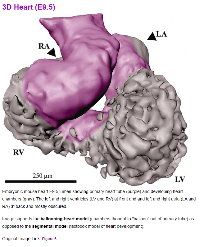
The challenge is to model the early development of the mouse heart from a simple tube to embryonic day 9.5. By this stage the tube has grown, twisted through 90 degrees, bent and started to balloon and form the atrial and ventricular surfaces. The deformations might arise from an evolving pattern of isotropic specified growth or might arise from anisotropic specified growth. It is not known.
In the wind-up session, groups will be invited to describe their successes and failures and there will be votes for the best, the wackiest and the worst models.

Surface fitted to 3D images of stage ED9.5. Growth starts with a simple tube. Image from [3] Click here for web page
Links
- Interactive 3D model of the mouse heart
- Illustrations of the developing mouse heart
- 'Three-dimensional reconstruction of gene expression patterns during cardiac development' by Alexandre T. Soufan , Jan M. Ruijter , Maurice J. B. van den Hoff , Piet A. J. de Boer , Jaco Hagoort and Antoon F. M. Moorman
- 'Oriented clonal cell growth in the developing mouse myocardium underlies cardiac morphogenesis' by Sigolène M. Meilhac, Milan Esner, Michel Kerszberg, Julie E. Moss, and Margaret E. Buckingham
- Cardiac laterality: The role of the homeobox transcription factor Pitx2 in cardiogenesis