Main Page: Difference between revisions
Jump to navigation
Jump to search
MORE
Have you seen the original paintings? Do they exist?.
No edit summary |
No edit summary |
||
| (76 intermediate revisions by 2 users not shown) | |||
| Line 1: | Line 1: | ||
=<span style="color:DarkGreen;">Computational biology | =<span style="color:DarkGreen;">Computational biology</span>= | ||
---- | ---- | ||
==<span style="color:DarkGreen;">Growing complex biological shapes from patterns of gene expression</span>== | ==<span style="color:DarkGreen;">[[Software#Quantitative understanding of growing shapes: GFtbox|<span style="color:Green;"> '''Growing''']] complex biological shapes from patterns of gene expression</span>== | ||
{| border="0" width=100% style="background-color:#000000;" | {| border="0" width=100% style="background-color:#000000;" | ||
|- | |- | ||
| Line 18: | Line 16: | ||
[[Image:LabelledCropped GPT Snapdragon 2010-000570-0002 triple.png|120px]] | [[Image:LabelledCropped GPT Snapdragon 2010-000570-0002 triple.png|120px]] | ||
|} | |} | ||
<br> | |||
[[Software#Quantitative understanding of growing shapes: GFtbox|<span style="color:Green;">'''MORE'''</span>]]<br> | |||
==<span style="color:DarkGreen;">[[Software#Viewing and measuring volume images: VolViewer|<span style="color:Green;"> '''Viewing''']] three dimensional volume (microscopy) images== | |||
{| border="0" width=100% style="background-color:#000000;" | {| border="0" width=100% style="background-color:#000000;" | ||
|- | |- | ||
| Line 41: | Line 38: | ||
[[Image:Arableaf_ath8_OPT.png|50px]] | [[Image:Arableaf_ath8_OPT.png|50px]] | ||
|} | |} | ||
<br> | |||
[[Software#Viewing and measuring volume images: VolViewer|<span style="color:Green;">'''MORE'''</span>]] | |||
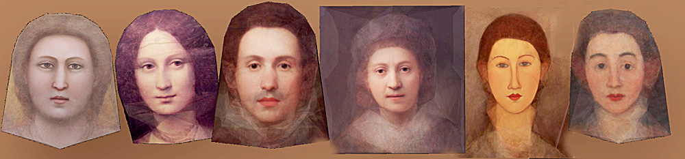
[[Software# | ==[[Software#Analysing shapes in 2D and 3D: AAMToolbox|<span style="color:Green;">'''Analysing'''</span>]] shapes: faces, leaves and flowers== | ||
{| border="0" width=100% style="background-color:#000000;" | |||
|- | |||
[[Image:PortraitsMEANSsmaller.jpg|800px]] | |||
|-} | |||
<br> | |||
[[Software#Analysing shapes in 2D and 3D: AAMToolbox|<span style="color:Green;">'''MORE'''</span>]]<br> | |||
Have you seen the original paintings? Do they exist?. <br><br> | |||
=<span style="color:Navy;">Algorithms= | |||
---- | |||
==[http://cmpdartsvr3.cmp.uea.ac.uk/wiki/BanghamLab/index.php/Software#MSERs.2C_extrema.2C_connected-set_filters_and_sieves <span style="color:Navy;">'''Vision''':] MSER's, extrema, filter-banks, Sieves and '''Scale-space'''== | |||
{| border="0" width=100% style="background-color:#ffffff;" | |||
|- | |||
|align="center"| | |||
[[Image:Cameraman_iso_topview.jpg|300px|AAMToolbox]] | |||
[[Image:Cameraman_iso_tree.jpg|300px|AAMToolbox]] | |||
|} | |||
=<span style="color:Navy;"> | [[Software#MSERs.2C_extrema.2C_connected-set_filters_and_sieves|<span style="color:Navy;">'''MORE'''</span>]] | ||
==[http://cmpdartsvr3.cmp.uea.ac.uk/wiki/BanghamLab/index.php/Software#Art.2C_extrema_of_light_and_shade:_PhotoArtMaster <span style="color:Navy;">'''Applications'''</span>]' <span style="color:Navy;">of non-linear filter banks (sieves) and the art of light and shade</span>== | |||
{| border="0" width=100% style="background-color:#ffffff;" | |||
|- | |||
|align="center"| | |||
[[Image:Colour_sieve.jpg|600px|AAMToolbox]] | |||
|} | |||
These images were produced from photographs using '''ArtMaster''' (formally known as '''PhotoArtMaster'''). The software received many favourable reviews when it was released (e.g. [http://graphicssoft.about.com/cs/photoart/gr/photoartmasterg.htm "This software can give you a lot of satisfaction from your everyday photos"], [http://graphicssoft.about.com/library/products/aafpr_photoartmaster1.htm] | |||
[http://cmpdartsvr3.cmp.uea.ac.uk/wiki/BanghamLab/index.php/Software#The_final_version_of_the_Windows_ArtMaster2.0_is_downloadable_here_with_no_support The final (so far unpublished) version of ArtMaster including code is downloadable from here.] I cannot provide support but quite of lot of documentation is available within [http://cmpdartsvr1.cmp.uea.ac.uk/downloads/software/SieveWebPages/a4a_2_screensize.pdf <span style="color: Chocolate">''''this document''''' </span>] | |||
[http://cmpdartsvr3.cmp.uea.ac.uk/wiki/BanghamLab/index.php/Software#Art.2C_extrema_of_light_and_shade:_PhotoArtMaster <span style="color:Navy;">'''MORE'''</span>] | |||
==[[Software#Reaction-diffusion and morphogenesis| <span style="color:Navy;"> '''Reaction-diffusion'''</span>]] <span style="color:Navy;">and morphogenesis - the growth of shapes== | |||
==<span style="color:Navy;">Reaction-diffusion and morphogenesis - the growth of shapes== | |||
{| border="0" width=100% style="background-color:#000000;" | {| border="0" width=100% style="background-color:#000000;" | ||
|- | |- | ||
| Line 63: | Line 82: | ||
[[Image:tentacles_morphogenesis.png|600px]] | [[Image:tentacles_morphogenesis.png|600px]] | ||
|} | |} | ||
This image forms part of a 'journey' in the Science Museum of London's 'Journeys of Invention' [http://www.sciencemuseum.org.uk/journeys iPad app.]<br><br> | |||
[[Software#Reaction-diffusion and morphogenesis|<span style="color:Navy;">'''MORE'''</span>]]<br><br> | |||
[[ | =[[Andrew personal | Andrew outside activities]]<br> | ||
Latest revision as of 13:03, 27 October 2014
Computational biology
Growing complex biological shapes from patterns of gene expression
|
|


Viewing three dimensional volume (microscopy) images
|
|


Analysing shapes: faces, leaves and flowers

MORE
Have you seen the original paintings? Do they exist?.
Algorithms
Vision: MSER's, extrema, filter-banks, Sieves and Scale-space
|
|


Applications' of non-linear filter banks (sieves) and the art of light and shade
|
|

These images were produced from photographs using ArtMaster (formally known as PhotoArtMaster). The software received many favourable reviews when it was released (e.g. "This software can give you a lot of satisfaction from your everyday photos", [1]
The final (so far unpublished) version of ArtMaster including code is downloadable from here. I cannot provide support but quite of lot of documentation is available within 'this document
Reaction-diffusion and morphogenesis - the growth of shapes
|
|


This image forms part of a 'journey' in the Science Museum of London's 'Journeys of Invention' iPad app.
MORE